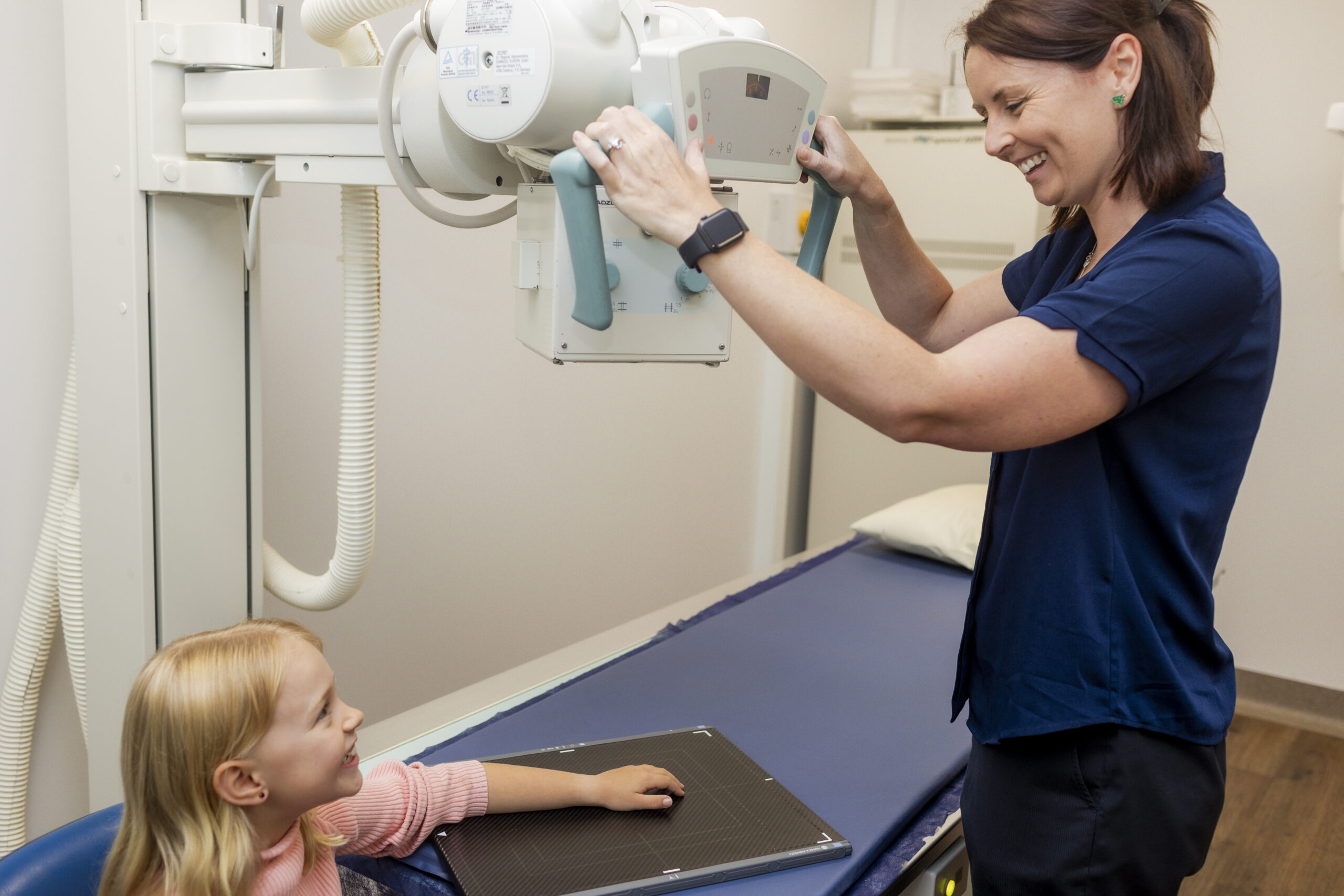
What is an X-Ray?
An X-ray is a powerful medical imaging technique that employs a small amount of x-ray energy to produce images of the inside of your body. Commonly used to visualise bones, X-rays can also help diagnose and monitor various conditions by giving insight into areas such as the lungs, chest, and abdomen. Doctors often refer patients for X-rays to diagnose fractures, detect infections, locate foreign objects, or monitor the progression of diseases.
What happens during an X-Ray?
An X-ray is a powerful medical imaging technique that employs a small amount of x-ray energy to produce images of the inside of your body. Commonly used to visualise bones, X-rays can also help diagnose and monitor various conditions by giving insight into areas such as the lungs, chest, and abdomen. Doctors often refer patients for X-rays to diagnose fractures, detect infections, locate foreign objects, or monitor the progression of diseases.

Our X-Ray Services
Dental X-Ray & OPG
A dental X-ray, including Orthopantomogram (OPG) scans, offers a detailed view of the mouth that goes beyond what can be seen with the naked eye. This imaging technique provides essential insights into the health of your teeth, jawbone, and surrounding oral structures. Whether it’s identifying cavities hiding between teeth, examining the roots of a troublesome tooth, or evaluating the jaw for orthodontic treatments, dental X-rays play a pivotal role in comprehensive oral care. Doctors and dentists recommend these images to ensure timely interventions and optimal dental health.
Chest X-Ray
A chest X-ray helps to visualise the heart, lungs and airways. It is often used to detect lung infections, most commonly pneumonia, leading to prompt and necessary treatment. Chest X-rays are also performed pre or post surgery. They can identify heart conditions, such as congestive heart failure, enlarged heart, or fluid around the heart and is a vital tool in diagnosing pneumothorax, a collapsed lung.
Chiropractic Spine X-Ray
If you are under the care of a chiropractor, they may refer you to have an X-ray of the spine to give them a better understanding of how they can help treat you. A spinal X-ray provides useful information relating to the vertebrae, discs and surrounding structures of the spine. It can help diagnose conditions such as curvatures of the spine, degenerative changes such as osteoarthritis, and can assess spinal alignment.
Knees, Hips & Shoulders X-Ray
X-ray is a useful diagnostic tool to visualise the bones that form joints in the body, big and small. It is often used to detect fractures and dislocations, forms of arthritis, and to assess and monitor joints pre and post joint replacements.
X-Ray and Radiology Bulk Billing
At GXU Radiology Specialists, all X-rays are bulk billed under Medicare with a referral from a GP or Specialist. While most X-rays referred from allied health professionals, such as physiotherapists and chiropractors are bulk billed, depending on what part of the body the X-ray is for, there may be a small fee associated with the imaging.
If you have a referral from an allied health professional for an X-ray, please let our reception team know, so that we can inform you of any potential fee.
Where we Operate our X-ray Services
Our X-rays are performed at our Thornton practice, conveniently situated within the Thornton Healthcare complex, which offers a range of medical services including pathology, dental, obstetrics, physiotherapy, and podiatry.
We are easily accessible, just a 30-minute drive from Newcastle and 10 minutes from Maitland, with the practice offering plenty of free onsite parking. We also regularly welcome patients from Taree, Gloucester, Scone, Tea Gardens, and the Central Coast.
If you’re taking public transport, a bus stop is conveniently located right outside our building on Anderson Drive. If you’re arriving by taxi, our friendly reception team can assist with arranging one for you.

Why Book an X-ray with GXU?
Founded in 2008, we have established ourselves as a trusted radiology provider.
Our team are passionate about radiology, and are committed to providing high-quality imaging for precise and accurate reports.
We are an accredited practice, affiliated with HDAA and the Royal Australian and New Zealand College of Radiologists, ensuring the highest standards of practice.
All reports are completed by specialists and made easily accessible, so your healthcare provider can receive timely, accurate insights.
We’re here when you need us most, offering same day appointments for urgent examinations.
Are There any Risks?
There’s no need to worry about the amount of radiation you receive from an X-ray. For example, a chest X-ray exposes you to a very small dose—about the same as the natural background radiation you’re exposed to over a few days of everyday life. Background radiation comes from natural sources like the sun, the soil, and even some foods we eat.
So, having a chest X-ray adds only a tiny amount to the radiation you’re already exposed to naturally. This minimal radiation helps doctors see inside your body to accurately diagnose and treat medical conditions. The benefits of having an X-ray far outweigh any minimal risks, so there’s no need to stress about the radiation involved.
What Our Customers Say
EXCELLENTTrustindex verifies that the original source of the review is Google. Was very happy with how professional everyone was. Dr was so gentle with cortisone injection. I found the staff be be polite and caring. Would highly recommendTrustindex verifies that the original source of the review is Google. Staff were very friendly and professional. My Biopsy was painless with every step explained. Could not have asked for better care. I highly recommend GXU.Trustindex verifies that the original source of the review is Google. I was running late and they still fit me in. Explained the procedure thoroughly. All staff were amazing. Thoroughly recommend.Trustindex verifies that the original source of the review is Google. The staff were great the waiting time was good I had a good experience there with no complaints at allTrustindex verifies that the original source of the review is Google. Greeted by friendly and professional staff who talked through the MRI process in detail and made it as comfortable as possible throughout.Trustindex verifies that the original source of the review is Google. Staff were friendly, polite and helpful. I had a ct scan guided injection in my back and the procedure went smoothly and was pain free. Highly recommendTrustindex verifies that the original source of the review is Google. Staff were courteous, professional and kind. It was an overall a good experience and I will definately go there again.Trustindex verifies that the original source of the review is Google. Friendly efficient, caring, they always go beyond to get you into an earlier appointment if they canTrustindex verifies that the original source of the review is Google. The experience attending this clinic is second to none. The care and treatment is patient centred. Everyone is so lovely, warm and welcoming from the moment you walk in the door. Best experience I have had attending a radiology clinic in years. Highly recommend 😊
X-Ray Appointments & Walk-Ins
We offer a walk-in service for x-ray and bookings are not required. However, you are welcome to book online if you would prefer by using the link below.
Frequently Asked Questions
How does an X-Ray work?
X-rays are a form of energy, much like visible light or microwaves, but with a much higher energy level. This high energy allows X-rays to pass through the body and produce detailed images. The images are created because different parts of the body absorb the X-rays in varying amounts. Dense structures like bone absorb more of the X-ray beam and appear light gray on the image. Softer, less dense tissues absorb less, appearing darker. These variations in absorption create a clear picture of the body’s internal structures. X-ray imaging is particularly good at assessing bones and looking for fractures.
How do I prepare for an X-ray?
Before having an X-ray, we recommend wearing comfortable, loose-fitting clothing. Depending on the area of the body being imaged, you might be asked to change into a gown. It’s important to remove all metallic objects, including jewellery, eyeglasses, and certain types of clothing fasteners, as these can interfere with the imaging. Inform the radiographer of any possibility of pregnancy or if you have any implanted medical devices.
How long does an X-ray take?
The process of acquiring the images takes a few seconds. Setting up and positioning takes several minutes. The overall duration of the examination depends on how many regions you are having imaged.
When can I expect the results of my X-rays?
The time that it takes your doctor to receive a written report on the test or procedure you have had will vary, depending on:
- the urgency with which the result is needed and the findings
- the complexity of the examination
- whether more information is needed from your doctor before the examination can be interpreted by the radiologist
- whether you have had previous x-rays or other medical imaging that needs to be compared with this new test or procedure (this is commonly the case if you have a disease or condition that is being followed to assess your progress)
- how the report is conveyed from the practice to your doctor (in other words, email, fax or mail)
Rest assured, if your doctor requires urgent results, we will prioritise and deliver them promptly.
Do you need a referral for an X-ray in NSW?
Yes, all X-rays performed require a referral from a GP, Specialist or allied health professional. A referral provides us with relevant information regarding your medical history and the clinical reason as to why you may need an X-ray.
Is an MRI more expensive than an X-ray?
Yes, an MRI is generally more expensive than an X-ray. In Australia, most X-ray procedures are covered by Medicare, making them affordable and widely accessible. X-rays are quick, usually taking only a few minutes.
MRI scans provide highly detailed images and can take up to 60 minutes. They require specialised radiographers and may not be fully covered by Medicare, so patients might need to pay some or all of the cost.
X-rays and MRIs offer different information. X-rays are excellent for imaging bones, while MRIs are superior for soft tissues. The choice between them depends on the specific medical information your doctor needs.


